收缩
Loading

Biotin anti-human CD11c;Clone#:3.9Host/Isotype:Mouse IgG1, κ   

详细描述

Clone: 3.9
Isotype: Mouse IgG1, κ
Workshop Number: III NL707
Reactivity: Human, Cross-Reactivity: Chimpanzee, Baboon, African Green, Cynomolgus, Rhesus, Squirrel Monkey
Formulation: Phosphate-buffered solution, pH 7.2, containing 0.09% sodium azide.
Preparation: The antibody was purified by affinity chromatography, and conjugated with biotin under optimal conditions. The solution is free of unconjugated biotin.
Concentration: 0.5 mg/ml
Storage & Handling: The antibody solution should be stored undiluted between 2°C and 8°C. Do not freeze.
Application:

FC - Quality tested

Application Notes:

Clone 3.9 preferentially binds the activated form of CD11c, is specific for I domain of CD11c, and is able to partially block the binding of CD11c and ICAM-4. 3.9 binding is divalent cation dependent.12 While analyzing blood, it is best to use heparin as the anti-coagulant and not EDTA. Since the ability of clone 3.9 to bind to its target is divalent cation dependent, the usage of EDTA as an anti-coagulant may be detrimental to staining due to its chelating properties.

Additional reported applications (for the relevant formats) include: immunohistochemical staining of acetone-fixed frozen tissue sections4, and functional assays5,6.

Recommended Usage:

Each lot of this antibody is quality control tested by immunofluorescent staining with flow cytometric analysis. For flow cytometric staining, the suggested use of this reagent is ≤2.0 µg per million cells in 100 µl volume. It is recommended that the reagent be titrated for optimal performance for each application.

Application References:

1. Schlossman S, et al. Eds. 1995. Leucocyte Typing V. Oxford University Press. New York.
2. Knapp W, et al. 1989. Leucocyte Typing IV Oxford University Press. New York.
3. McMichael A, et al. Eds. 1987. Leucocyte Typing III Oxford University Press. New York.
4. Vainer B, et al. 2000. Am. J. Surg. Pathol. 24:1115. (IHC)
5. Ottonello L, et al. 1999. Blood 93:3505.
6. Metelitsa LS, et al. 2002. Blood 99:4166.
7. Sadhu C, et al. 2007. J. Leukoc. Biol. doi:10.1189/jlb.1106680. PubMed
8. Ihanus E, et al. 2007. Blood 109:802-810.
9. Gurer C, et al. 2008. Blood 112:1231. PubMed
10. Asai A, et al. 2009. J. Lipid Res. 50:95. PubMed
11. Yoshino N, et al. 2000. Exp. Anim. (Tokyo) 49:97. (FC)
12. Sadhu C, et al. 2008. J. Immunoass. Immunoch. 29:42. (FC)

Human peripheral blood granulocytes stained

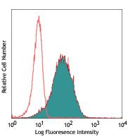
Human peripheral blood granulocytes stained with biotinylated 3.9, followed by Sav-PE

Description:

CD11c is a 145-150 kD type I transmembrane glycoprotein also known as integrin αX and CR4. CD11c non-covalently associates with integrin β2 (CD18) and is expressed on monocytes/macrophages, dendritic cells, granulocytes, NK cells, and subsets of T and B cells. CD11c has been reported to play a role in adhesion and CTL killing through its interactions with fibrinogen, CD54, and iC3b.

Other Names: Integrin αX subunit, CR4, p150, ITGAX
Structure: Integrin, type I transmembrane glycoprotein, associates with integrin β2 (CD18), 145-150 kD
Distribution: Myeloid, dendritic cells, NK cells, B cells and T cell subsets
Function: Adhesion, CTL killing
Ligand Receptor: CD54, fibrinogen, iC3b, ICAM-1, ICAM-4
Antigen References:

1. Petty H. 1996. Immunol. Today 17:209.
2. Springer T. 1994. Cell 76:301.
3. Ihanus E, et al. 2007. Blood 109:802-810.


  • 目录号:2108055
  • 品牌:Sony Biotechnology
  • 规格:25μg
  • 目录价:¥2064.00
  • 市场价格:¥1341.60
  • 会员价格:¥1279.68
  • 数量:

购买 发起团购

结束时间:
参加人数:人