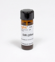
IVISENSE DIR 750 FLUOR CELL LABEL DYE 现货推荐!!

详细描述

XenoLight DiR (DiIC18(7) or 1,1’-dioctadecyltetramethyl indotricarbocyanine Iodide)
Absorption/Emission: 748/780 nm
Ideal IVIS filter set: 710 ex/760 em
XenoLight DiR is a lipophilic, near infra-red (NIR) fluorescent cyanine dye ideal for staining cytoplasmic membrane. The two long 18-carbon chains insert into the cell membrane, resulting in specific and stable cell staining with negligible dye transfer between cells.
XenoLight DiR NIR Fluorescent Dye in combination with PerkinElmer’s IVIS imaging systems can be used for non-invasive imaging of T-cell or stem cell homing in vivo . The near infrared property of this dye makes it ideal for in vivo imaging because of significantly reduced autofluorescence from the animal at higher wavelengths.
“For laboratory use only. This product is intended for animal research only and not for use in humans.”
Cell Staining
XenoLight DiR can be applied to fluorescence staining of primary cells, such as embryonic stem cells, bone marrow derived stem cells, adipose derived stem cells, lymphocytes and erythrocytes, which will enable fluorescence detection of stained cells and their in vivo distribution. Since DiR has excitation and emission maxima in the NIR range, fluorescence detection of DiR stained cells will have less interference from autofluorescent tissue background, resulting in high sensitivity of detection.
In vivo Imaging of DiR stained Spleen T-cells distribution

XenoLight DiR stock was prepared by suspending 25mg in 3 ml ethanol. Working solution of 320 ug/ml was prepared by diluting 199 ul of stock solution in 5 ml PBS. T-cells isolated from the spleen were incubated with 320 ug/ml DiR. After 30 min incubation, cells were spun down for 3 min at 1000rpm at 4ºC resulting in a blue pellet. Cells were washed twice in PBS and injected intravenously (i.v.) (5.0E+06 cells/mouse). Control group was injected with 5.0E+06 cells/mouse in PBS. Mice were imaged with IVIS Spectrum at 10 min, 1hr, 6hr and 24 hrs post injection. Ideal filter set for DiR imaging is 710 nm excitation and 780 nm emission. Mice were imaged dorsally as well as ventrally at all time points. Brain, bones, spleen, liver, lungs and kidneys were harvested for ex vivo imaging 24 hrs post injection.
Non-invasive in vivo imaging showed the homing process of injected T cells to the liver and spleen in real time, which was confirmed by ex vivo imaging.

